Something very similar to Pemphigus Vulgaris is Pemphigoid. Mucous Membrane Pemphigoid is a chronic autoimmune mucocutaneous vesiculobullous disease where autoantibodies target components of the basement membrane.
Don’t have time to read this article? We get it. Download the Diagnosing Vesicular Ulcerative Conditions checklist to get the key information and images from this article plus all the other conditions we cover in the Dentist’s Guide to Oral Pathology.

Mucous Membrane Pemphigoid Diagnosis
Where do we find Mucous Membrane Pemphigoid (MMP) in patients?
Blistering lesions that primarily affect the various mucous membranes of the body. The mucous membranes of the mouth and eyes are most often affected.
They can occur anywhere: the oral cavity, eyes, vaginal canal, or esophagus. Anywhere can be involved. However, you may or may not see vesicles or bullae and then erosive as well. This is secondary to autoantibodies that are attacking the basement membrane.
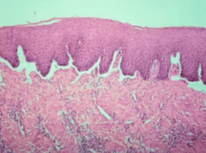
Instead of the desmosome attachments between cells, we’ve got the hemidesmosomes that are involved.
You can see a nice clean separation – you’re not seeing those basal cells. The basal cells are still attached to the epithelium and this is where the separation is occurring; it’s subepithelial.
Clinical Features of Mucous Membrane Pemphigoid
- Adults > 40
- F > M
- No ethnic predilection
- Painful, +/- vesicles
- Predilection for gingiva
- Eyes
- Biopsy needed for definitive differential
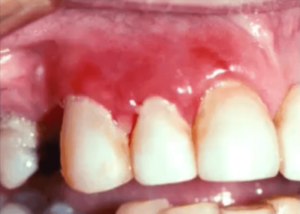
Here’s a clinical picture of pemphigoid.
So you’re not gonna be able to tell the difference clinically. You may or may not see vesicles, but widespread ulcerative lesions here involving the gingiva.
Oftentimes these patients are seeing or have seen multiple clinicians and they still have the same presentation. A lot of dentists, in general, think of gingivitis. Not surprisingly because there’s a lot of calculus here. So the first thing you might do here is clean the patient’s teeth, but if the lesions don’t resolve you’ve got to start thinking outside the box.
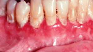
These lesions are erosive lesions – they’re not secondary to the plaque. It is so painful for this patient to brush that they don’t brush.
As you can imagine, if you’re brushing on your connective tissue and nothing is protecting your connective tissue, it would be extremely painful. This is why they have so much plaque accumulation which exacerbates the condition. It is so important to maintain oral hygiene.
Ocular Mucous Membrane Pemphigoid
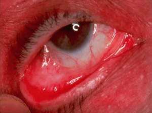
When the mucous membrane of the eye is traumatized it will heal right on top of the eyeball, or the iris, and that is called a symblepharon, a mucosal attachment to the eye.
When this becomes widespread, it can cause blindness.
If you ever have a patient that has been diagnosed with pemphigoid, you need to refer them to an ophthalmologist as soon as possible in order for the ophthalmologist to evaluate and treat as necessary.