In this article we’ll explore three primary tests to help diagnose temporomandibular disorders:
- Imaging tests
- Physical medicine tests
- Serologic tests
1. Imaging Tests
A. Panoramic Radiographs
A panoramic radiograph is utilized primarily to have a general vision of the maxillaries. For the TMJ, it is possible to do an initial evaluation of the condylar shape, presence of subchondral erosion, cysts, flattening and erosion.
The clinical history and/or examination finding needed to order this diagnostic test includes:
- Traumatic injury to the jaw
- Presence of asymmetry of the mandible with the midline or chin being off-center
- Severe pain and/or crepitation of the joint on motion
- Suspected maxillary sinus pathology
Related Reading:
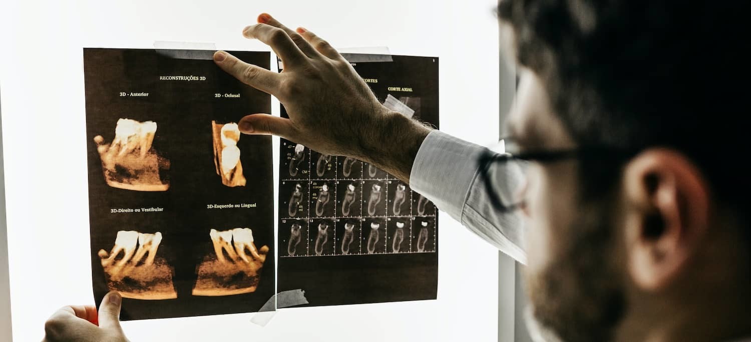
B. Medical (Spiral) CT or Cone Bean CT (CBCT) of Jaw and TMJ
A computerized tomographic image (CT) mostly utilized to evaluate a bony or soft tissue lesions. Some examples are growths, tumors, and bone infections or reaction in the maxilla or mandible.
Compared to a CBCT a medical CT has a relatively higher radiation dose. Either image is utilized when you need a better osseous image of a jaw structure, for example, the TMJ or any region of the mandible or maxilla where pathology is suspected.
While the estimates of radiation dose vary, the CBCT has a radiation dose equivalent to 2-3 panoramic radiographs and medical CT is 5-10x more radiation than a CBCT (depending on the type of equipment).
The clinical history and/or examination finding needed to order this diagnostic test includes:
- Suspected sinus obstructions based on complaint of reduced nasal airflow
- Suspected altered jaw or facial bone structure (facial or mandibular asymmetry)
- Suspected tumor of the orofacial region
- Suspected traumatic injury to the jaw and/or maxillofacial region
- Any type of arthritic disease affecting the jaw system
C. MRI Imaging of the TM Joint
An MRI is indicated for the TMJ when an image of the disc is required. Usually as a baseline or as a follow up for a DDNR. It is the image of choice with there is an acute onset loss of motion of the jaw, prior to decide on a major occlusal therapy.
The clinical history and/or examination finding needed to order this diagnostic test includes:
- Recent onset of jaw opening limitation (<35mm)
- Pain in the affected TMJ with opening
- Joint noises in the TMJ that stopped with recent loss of opening ability.
Like what you’re learning? Explore our online, postgraduate Orofacial Pain and Oral Medicine degrees.
2. Physical Medicine Tests
A. Passive Stretch Test (With & Without Vapocollant Spray)
A strategy to differentiate between a muscle and joint-related reduced range of motion of the jaw is to use passive stretching. If full opening (>45mm) is easily achieved with vapocoolant spray, then the disk is not displaced and you are more likely dealing with a trismus or spasm. Once the diagnosis is achieved, the stretching procedure can be done daily to recover the full range of motion. The test is performed by stretching open the jaw with mild to moderate force between the maxillary and mandibular teeth.
The clinical history and/or examination finding needed to order this diagnostic test includes:
- Inability to perform a wide opening of the jaw
- Unassisted opening (a.k.a. active opening) of the jaw is <35mm of interincisal distance
- Opening is moderately painful
B. Stretch Localization Test
In order to locate the possible site of restriction, the clinician will perform a manual stretch of the jaw, and ask the patient to point where is the pain. The patient will point to the structure or region that produces pain (for example, anterior portion of masseter muscle or anterior fibers of temporalis).
The clinical history and/or examination finding needed to order this diagnostic test includes:
- Limitation of active opening of the jaw
- Traumatic onset of the limitation
- Suspected focal myositis or fibrosis of the muscle
Related Reading: Trigger Point Mapping: Theory & Step-by-Step Technique
3. Serologic Testing
A. Autoimmunity Panel
Diseases such as rheumatoid arthritis, systemic lupus, Sjogren’s disease, myositis, and mixed connective tissue disease will be identified using serologic techniques.
The clinical history and/or examination finding needed to order this diagnostic test includes:
- Multiple sites of pain and soreness suggestive of an autoimmune disease such as lupus
- Swollen and diminished function of the salivary glands suggestive of Sjogren’s Syndrome
- Facial numbness and pain of unknown origin (without local causative factors evident)
Earn a Master’s Degree in Orofacial Pain and Oral Medicine Online
Like what you’re learning? Consider enrolling in the Herman Ostrow School of Dentistry of USC’s online, competency-based 1-year certificate program or 3-year Master’s program in Orofacial Pain and Oral Medicine.
Disclaimer
The information and resources contained on this website are for informational purposes only and are not intended to assess, diagnose, or treat any medical and/or mental health disease or condition. The use of this website does not imply nor establish any type of provider-client relationship. Furthermore, the information obtained from this site should not be considered a substitute for a thorough medical and/or mental health evaluation by an appropriately credentialed and licensed professional. Commercial supporters are not involved in the content development or editorial process.